Catheter Mounts and Closed Suction
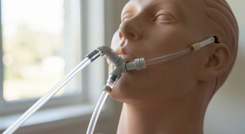
Between the breathing circuit and the patient's airway sits a short piece of flexible tubing called a catheter mount. It looks like a minor part, but it is the link that absorbs the weight and movement of the whole circuit so that none of it pulls on the endotracheal tube. It is also the point where suction is connected, and where a closed suction system keeps the patient on the ventilator while secretions are cleared. This guide explains how a catheter mount is built and how closed suction works.
What does a catheter mount do?
A catheter mount connects the Y-piece of the breathing circuit to the airway device, adding a flexible length that lets the circuit move without transmitting that movement to the tube. Because the tube sits in the trachea, any pull on it is uncomfortable and can displace it, so the mount acts as a shock absorber. It also provides a port for a suction catheter and a place to take gas samples. A well chosen mount is light, short enough not to add much dead space, and flexible enough to take up the circuit's movement.
Why does a double swivel matter?
A swivel elbow lets the mount rotate, and a double swivel lets it rotate in two planes at once. That matters when a patient turns their head or when the circuit is repositioned, because without a swivel the movement becomes torque on the tube. The double swivel also makes it easier to keep the assembly oriented so that the circuit is supported and does not drag. When the mount is rigid or the swivel is stiff, the strain is passed to the airway, which is exactly what the mount is meant to prevent.
How does closed suction work?
In closed suction, a catheter is enclosed in a sterile sleeve attached to the circuit, so it can be advanced into the airway and withdrawn without disconnecting the patient from the ventilator. The sleeve keeps the catheter clean between uses and lets suction be applied while ventilation continues, which avoids the loss of pressure and the risk of contamination that come with an open system. The catheter is advanced to the measured depth, suction is applied briefly as it is withdrawn, and the catheter is pulled back into its sleeve. Because the circuit stays closed, the patient does not lose positive end expiratory pressure or oxygen during the procedure.
How is the assembly kept light?
Every component between the Y-piece and the airway adds weight and dead space, and the patient feels both. A catheter mount, a closed suction unit, a filter or an HME and the swivel elbows together form a stack that can become heavy enough to pull on the tube. The practical approach is to keep the assembly as short as the clinical need allows, to support the circuit so its weight rests on the bed rather than the airway, and to choose mounts and connectors that are light. The ventilator breathing circuits guide explains why dead space matters most in small patients, where the same stack is a larger fraction of each breath.
Where does the filter or HME sit?
On a circuit with closed suction, a filter or heat and moisture exchanger often sits between the Y-piece and the catheter mount, or between the mount and the airway, depending on the setup. Each position has consequences: a device closer to the patient conditions the gas at the airway but adds dead space there, while a device further from the patient protects more of the circuit. The bacteria and viral filters guide and the HME filters guide set out the trade offs. Whatever the order, the whole stack is planned as one assembly rather than assembled part by part.
How the suction catheter is sized
A closed suction catheter is chosen so that its outer diameter is no more than about half the inner diameter of the airway tube. A catheter that is too large blocks the tube as it is advanced, so the patient cannot breathe around it, and suction then removes gas from the lung as well as secretions. A catheter that is too small clears secretions poorly. The correct depth is set from the length of the airway device, and it is marked so that the catheter is not advanced beyond the carina. Because the catheter sits inside a sleeve, its size is confirmed before it is fitted, and the depth is checked at the same time. Matching the catheter to the tube, and to the patient's secretions, is what makes closed suction effective rather than merely routine.
What to check before and after suction
Before suction, confirm the catheter moves freely in its sleeve, the suction pressure is set as policy requires, and the depth to advance the catheter is known. Check the mount and swivel are intact and the assembly is supported. During suction, watch the patient's oxygen saturation and heart rate, keep the suction brief, and stop if the patient deteriorates. After suction, confirm the catheter is fully withdrawn into the sleeve, the circuit is still sealed and the ventilator readings are back to their previous values. A short and repeatable routine keeps closed suction safe and keeps the airway protected from the weight and movement of the circuit around it.